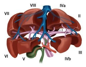
The functional subdivision of the liver is based on the portal branching into individual, mutually independent subunits, the segments.
Usually, the portal vein divides at the hepatic hilum into a right and left main trunk. The boundary of these supply territories lies at the cava–gallbladder line ("Cantlie line"). Through a further bifurcation of the respective portal vein trunk, an anteromedial and a posterolateral trunk arise on the right side for liver segments V/VIII and VI/VII, respectively. The left main trunk runs transversely to the left and then, as the pars umbilicalis, anteriorly, and terminates at the insertion site of the ligamentum teres hepatis in the so-called recessus of Rex. The left portal main trunk gives off branches for the two left-lateral segments II and III as well as for the medial segments IVa and IVb. The lobus caudatus holds a special position, as it may receive substantial inflow from both the left and the right portal main trunk.
According to Couinaud, eight portal-venous liver segments are distinguished, which, beginning with the lobus caudatus as segment I, are numbered clockwise:
Segment I………………………..Lobus caudatus
Segments I/II/III………………….lateral left liver lobe
Segment IV………………………left paramedian sector (lobus quadratus)
Segments I/II/III/IV………………left half of the liver
Segments V/VIII…………………right paramedian sector
Segments VI/VII…………………right lateral sector
Segments V/VI/VII/VIII…………..right half of the liver
The liver is traversed in a caudocranial direction by three main venous trunks, namely the right, middle, and left hepatic veins, which subdivide the liver into a total of four hepatic sectors. The left hepatic vein drains almost exclusively the left-lateral liver lobe and usually unites, shortly before its entry into the vena cava, with the middle hepatic vein, which runs along the cava–gallbladder line. The right hepatic vein runs between the posterolateral and anteromedial segments. The lobus caudatus has an independent venous outflow, consisting of multiple small veins that drain dorsally directly into the vena cava, the so-called Spigelian veins.
In fact, the regularity of vascular branching described by Couinaud is present only in the rarest of cases; a multitude of branching variants exists, with the size of the supply territories being variable in individual cases.
The portal hila of liver segments II, III, and IV lie extrahepatically and can be dissected out relatively easily in the anterior portion of the left umbilical fissure. The hila of the right-sided liver segments lie intrahepatically; exceptions occur occasionally and mostly concern segment VI. Even more variable than the anatomy of the portal vein is that of the hepatic veins.