
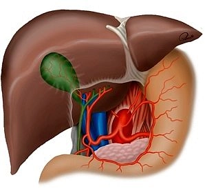
The pancreas, composed of lobules, has a reddish-grey colour, is 14 – 18 cm long, and weighs 65 – 80 grams. It is located at the level of the 1st and 2nd lumbar vertebrae and extends in a wedge-shaped manner from the epigastric region into the left hypochondriac region. Owing to its developmental history, the organ is in close proximity to the upper abdominal organs and vessels.
The pancreas is encased in capsule-like connective or fatty tissue and is divided into three sections: caput, corpus, and cauda. While a somewhat firmer connective tissue plate is located in the posterior head region, the gland is otherwise predominantly loosely connected dorsally with the connective tissue. As a retroperitoneal organ, the gland is covered on its anterior surface with peritoneum.
The widest part of the gland is the caput pancreatis, which, located to the right of the spine, fits into the loop formed by the duodenum. Both the anterior and posterior surfaces of the duodenum can be overlapped by glandular tissue to varying extents here. The caput encompasses the superior mesenteric vein from behind with its caudal portion (processus uncinatus), occasionally also the artery. The groove located in the processus uncinatus and the rest of the pancreatic head is referred to as the incisura pancreatis.
The part of the pancreas located at the level of the 1st lumbar vertebral body, with a width of about 2 cm, represents the transition area from the head to the body and lies over the superior mesenteric vessels. From a surgical perspective, this section is also referred to as the neck of the pancreas.
The elongated body of the pancreas runs obliquely over the 1st and 2nd lumbar vertebrae upwards, bulges ventrally into the omental bursa, and arches towards the splenic hilum, with the transition into the tail occurring without precise anatomical demarcation. Dorsal to the body, alongside the spine, are the aorta, inferior vena cava, and superior mesenteric artery and vein.
The tail of the pancreas forms the pointed continuation of the glandular body and extends to or into the splenorenal ligament.
The pancreas can be configured in various shape variants, obliquely, S-shaped, transversely, and L-shaped. A horseshoe shape and an inverted V-shape have also been described. The transitions between the shape variants are fluid.