Start your free 3-day trial — no credit card required, full access included

Duodenohemipancreatectomy with Blumgart anastomosis and biliopancreatic separation

  1. Laparotomy

    Laparotomy
    Soundsettings

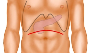
    The approach is a curved, cranially convex, transverse upper abdominal laparotomy. Subsequently, the exploration phase involves inspection and palpation of the entire abdomen to exclude distant metastases and signs of local unresectability.

    After inserting a sheet as abdominal wall protection, a pulley hook system is used.

    Note:

    Alternatively, in cases of a very acute costal angle, a median laparotomy may be performed.
  2. Opening of the Omental Bursa

    Opening of the Omental Bursa
    Soundsettings

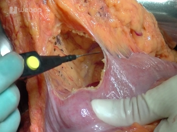
    Entering the omental bursa by detaching the omentum from the right colonic flexure to the left third of the transverse colon. Now the pancreas can also be well explored towards the left side, and it can be determined whether there is infiltration of the stomach or postpyloric duodenum. Adhesions between the pancreas and the posterior gastric wall are released.

    Note:

    The duodenum should be preserved over a distance of 2 to 3 cm. If this is not possible, the stomach must be partially resected.

  3. Kocher Manoeuvre

    Kocher Manoeuvre
    Soundsettings

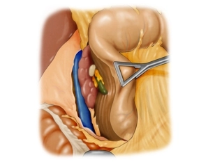
    First, detach the right colonic flexure from the anterior surface of the duodenum and the head of the pancreas. Then, the Kocher's mobilization of the duodenum is performed. The mobilized section of the colon is displaced to the left side, and the duodenum, following incision along its lateral edge, is freed together with the head of the pancreas from its retroperitoneal connections. The mobilization of the pancreatic head is continued beyond the aortic plane up to the ligament of Treitz until the pancreatic head can be completely encompassed from the dorsal side.

    The palpation of the mobilized pancreatic head is performed to exclude retroperitoneal infiltration, as well as the palpation of the lymphatic drainage pathways and the major vessels.

  4. Cholecystectomy

    Cholecystectomy
    Soundsettings

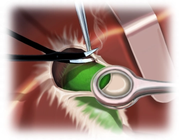
    The resection phase begins with the cholecystectomy: grasping the gallbladder and performing antegrade subserosal dissection from the liver bed using bipolar scissors, exposing the cystic duct and cystic artery, which are initially left on the subsequent specimen.

    Note:

    To prevent ascending cholangitis after biliodigestive anastomosis, cholecystectomy is mandatory for functional reasons during pancreatic head resection.

  5. Preparation of the Hepatoduodenal Ligament with Division of the Right Gastric Artery

    Preparation of the Hepatoduodenal Ligament with Division of the Right Gastric Artery
    Soundsettings

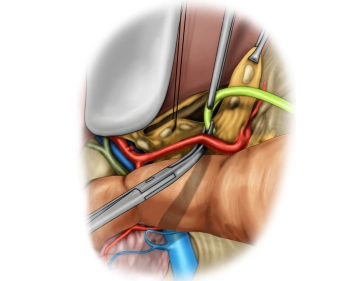
    The preparation within the hepatoduodenal ligament includes the exposure of the common hepatic artery, the proper hepatic artery, the gastroduodenal artery, the portal vein and the common bile duct (CBD).

    In this case, an anatomical variant of the liver's vascular supply is present. An atypical right hepatic artery arising from the superior mesenteric artery crosses the CBD dorsally.

    In this case, lymphadenectomy is performed along the left hepatic artery centrally towards the coeliac trunk. During this, the origin of the gastroduodenal artery is exposed.

    The division of the right gastric artery between ligatures marks the beginning of the mobilisation of the pylorus.

    Prior to this, the preparation of the ligament is completed with the exposure of the suprapancreatic portal vein up to behind the pancreas.

  6. Transection of the Postpyloric Duodenum and the Gastroduodenal Artery

    Soundsettings

    Now dissection and then transection of the duodenum with the stapler 3 cm postpylorically. Subsequently, transection first of the gastroepiploic arcade and then of the gastroduodenal artery. The central stump is managed with a transfixion ligature.

    Tip:

    Before transecting the gastroduodenal artery, trial clamping is advisable to rule out stenosis of the coeliac trunk.

Division and Skeletonisation of the First Jejunal Loop

Locate the first jejunal loop aboral to the ligament of Treitz. Taking the supplying vascular arcad

Activate now and continue learning straight away.

Single Access

Activation of this course for 3 days.

US$9.27 inclusive VAT
Add to cart

Most popular offer

webop - Savings Flex

Combine our learning modules flexibly and save up to 50%.

from US$7.20 / module

US$86.41/ yearly payment

price overview

general and visceral surgery

Unlock all courses in this module.

US$14.40 / month

US$172.83 / yearly payment

Add to cart
to top