Start your free 3-day trial — no credit card required, full access included

Duodenohemipancreatectomy with Blumgart anastomosis and biliopancreatic separation

Reading time readingtime 32:14 min.
  1. Laparotomy

    Laparotomy
    Soundsettings

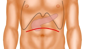
    The approach is a curved, cranially convex, transverse upper abdominal laparotomy. Subsequently, the exploration phase involves inspection and palpation of the entire abdomen to exclude distant metastases and signs of local unresectability.

    After inserting a sheet as abdominal wall protection, a pulley hook system is used.

    Note:

    Alternatively, in cases of a very acute costal angle, a median laparotomy may be performed.
  2. Opening of the Omental Bursa

    Opening of the Omental Bursa
    Soundsettings

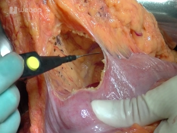
    Entering the omental bursa by detaching the omentum from the right colonic flexure to the left third of the transverse colon. Now the pancreas can also be well explored towards the left side, and it can be determined whether there is infiltration of the stomach or postpyloric duodenum. Adhesions between the pancreas and the posterior gastric wall are released.

    Note:

    The duodenum should be preserved over a distance of 2 to 3 cm. If this is not possible, the stomach must be partially resected.

  3. Kocher Manoeuvre

    Kocher Manoeuvre
    Soundsettings

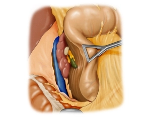
    First, detach the right colonic flexure from the anterior surface of the duodenum and the head of the pancreas. Then, the Kocher's mobilization of the duodenum is performed. The mobilized section of the colon is displaced to the left side, and the duodenum, following incision along its lateral edge, is freed together with the head of the pancreas from its retroperitoneal connections. The mobilization of the pancreatic head is continued beyond the aortic plane up to the ligament of Treitz until the pancreatic head can be completely encompassed from the dorsal side.

    The palpation of the mobilized pancreatic head is performed to exclude retroperitoneal infiltration, as well as the palpation of the lymphatic drainage pathways and the major vessels.

  4. Cholecystectomy

    Cholecystectomy
    Soundsettings

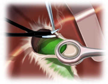
    The resection phase begins with the cholecystectomy: grasping the gallbladder and performing antegrade subserosal dissection from the liver bed using bipolar scissors, exposing the cystic duct and cystic artery, which are initially left on the subsequent specimen.

    Note:

    To prevent ascending cholangitis after biliodigestive anastomosis, cholecystectomy is mandatory for functional reasons during pancreatic head resection.

  5. Preparation of the Hepatoduodenal Ligament with Division of the Right Gastric Artery

    Preparation of the Hepatoduodenal Ligament with Division of the Right Gastric Artery
    Soundsettings

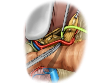
    The preparation within the hepatoduodenal ligament includes the exposure of the common hepatic artery, the proper hepatic artery, the gastroduodenal artery, the portal vein and the common bile duct (CBD).

    In this case, an anatomical variant of the liver's vascular supply is present. An atypical right hepatic artery arising from the superior mesenteric artery crosses the CBD dorsally.

    In this case, lymphadenectomy is performed along the left hepatic artery centrally towards the coeliac trunk. During this, the origin of the gastroduodenal artery is exposed.

    The division of the right gastric artery between ligatures marks the beginning of the mobilisation of the pylorus.

    Prior to this, the preparation of the ligament is completed with the exposure of the suprapancreatic portal vein up to behind the pancreas.

  6. Transection of the Postpyloric Duodenum and the Gastroduodenal Artery

    Soundsettings

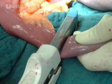
    Now dissection and then transection of the duodenum with the stapler 3 cm postpylorically. Subsequently, transection first of the gastroepiploic arcade and then of the gastroduodenal artery. The central stump is managed with a transfixion ligature.

    Tip:

    Before transecting the gastroduodenal artery, trial clamping is advisable to rule out stenosis of the coeliac trunk.

  7. Division and Skeletonisation of the First Jejunal Loop

    Division and Skeletonisation of the First Jejunal Loop
    Soundsettings

    Locate the first jejunal loop aboral to the ligament of Treitz. Taking the supplying vascular arcades into account, the jejunum is then transected with the stapler. Subsequently, the mesentery of the oral limb is dissected close to the intestine up to the duodenojejunal junction at the level of the ligament of Treitz.

    Note:

    In the demonstrated case, the dissection of the mesentery is performed between ligatures; this is considerably easier and faster with the aid of a sealing instrument based on diathermy or ultrasound.

  8. Passage of the Jejunal Stump / Preparation of the Superior Mesenteric Vein

    Soundsettings

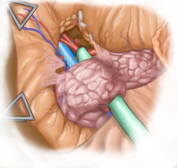
    The prepared jejunal stump is relocated through the mesocolon into the upper abdomen. Subsequently, the mesenteric vein is located at the lower edge of the pancreatic head. The superior mesenteric vein is traced beneath the pancreas.

    Note:

    In cases of unclear resectability, the preparation of the superior mesenteric vein must be prioritised, as this is the critical point for assessing the feasibility of a meaningful resection.

  9. Transection of the Pancreas and Common Bile Duct

    Transection of the Pancreas and Common Bile Duct
    Soundsettings

    Above this vessel, the pancreas is gently lifted and tunneled. Then the pancreas is passed under with an Overholt clamp and looped. This loop protects the superior mesenteric vein during the subsequent transection of the pancreas with a scalpel. Bleeding vessels at the distal resection surface are controlled with transfixion ligatures (4-0 monofilament, slowly absorbable). Then, further preparation of the pancreatic stump is performed in preparation for the anastomosis.

    Now, the common bile duct is closed distally with a suture and then transected proximal to the cystic duct junction.

    A swab is taken. Towards the liver, the bile duct is temporarily closed with a bulldog clamp.

    Tip:

    For reasons of blood supply, the common hepatic duct should be transected as close to the hilum as possible, proximal to the cystic duct junction.

  10. Detachment of the Pancreatic Head

    Detachment of the Pancreatic Head
    Soundsettings

    The complete detachment of the pancreatic head is performed by transecting the supplying portal vein branches.

    This completes the resection phase, and the specimen is sent for histopathological examination.

    An intraoperative frozen section examination of the pancreatic resection margin should confirm tumour-free resection margins. If portions of an invasively growing carcinoma or areas with high-grade epithelial dysplasia are still detectable at the resection margin, further resection up to a total pancreatectomy is indicated.

    Note:

    At the retroperitoneal dorsal resection margin, careful preparation is essential, as advanced tumour infiltration (R1 situation) is most likely to be expected in this area.

  11. Sealing of the Retroperitoneal Resection Surface

    Soundsettings

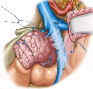
    Application of a powdered haemostatic agent to the retroperitoneal resection surface between the portal vein and the vena cava to reduce the risk of postoperative bleeding.

    For maximum hemostatic effect, it is important that blood clots are carefully removed, as the powder particles need direct contact with the tissue.

    Now, the powder is applied directly to the entire surface, and the edges must also be covered. It is not the thickness but the uniform coverage that is crucial.

    For approximately 2 minutes, uniform pressure is applied to the entire wound surface with one or two dry gauzes.

    Subsequently, irrigation with physiological saline solution is performed to release the gauze or to prevent adhesions.

  12. Oversewing and Retrocolic Elevation of the Jejunal Stump

    Soundsettings

    The aboral jejunal stump is oversewn and then guided retrocolically through a newly created mesocolic window into the right upper abdomen. It should come to lie tension-free in front of the pancreatic remnant.

  13. Pancreaticojejunostomy according to Blumgart I (Posterior Wall)

    Soundsettings

    The pancreaticojejunostomy is constructed in two layers with a duct-to-mucosa anastomosis in an end-to-side fashion. For this, the pancreas to the left of the superior mesenteric vein must be sufficiently mobilized and the duct probed. First, transpancreatic full-thickness U-sutures are placed through the pancreas to the serosa of the jejunal stump using a double-armed suture.

    After a pinpoint opening of the jejunum, the mucosa is first sutured up with 3 stitches. The inner suture line (duct-to-mucosa) is initially placed with double-armed sutures. The suture captures the pancreatic duct and the corresponding jejunum as a full-thickness suture. The direction of each needle is from inside to outside, so that the knots will later come to lie on the outside. Then the pre-placed transpancreatic U-sutures are tightened, thereby approximating the pancreatic stump to the jejunum. Subsequently, these sutures are tied without cutting the suture ends.

    With the tying of the duct-to-mucosa sutures, the posterior wall suture is then complete.

    Note:

    The main focus in the pancreatic anastomosis should be on tension-free adaptation and good perfusion conditions; obstruction of the pancreatic duct should be avoided.

  14. Pancreaticojejunostomy according to Blumgart II (Anterior Wall)

    Soundsettings

    The anterior wall suture is performed in the same manner: first the inner suture row with a duct-to-mucosa suture, and finally, using the already placed transfixing pancreatic suture, grasping the jejunal serosa on the opposite side, so that tying results in a telescopic invagination of the pancreatic stump into the jejunal stump.

  15. Formation of the Roux-Y Loop

    Soundsettings

    First, fixation of the jejunal loop carrying the pancreatic anastomosis in the mesocolic slit.

    In the next step, the bile duct anastomosis is created as an end-to-side anastomosis (hepaticojejunostomy) into a second jejunal loop formed according to Roux-Y.

    For this, a jejunal loop is first selected approximately 30 cm aboral to the mesocolic passage under transillumination of the vascular arcades. Avascular parts of the mesentery are transected with the cautery, and the crossing vessels are divided between clamps. Transection of the intestine with the stapler. The staple suture line of the efferent loop is oversewn in an invaginating manner.

    The efferent jejunal loop is transposed antecolically into the upper abdomen and approximated to the bile duct stump.

  16. Biliodigestive Anastomosis

    Soundsettings

    Pinpoint antimesenteric opening of the elevated jejunal stump and elevation of the mucosa with 3 everting interrupted sutures. Then hepaticojejunostomy as an end-to-side anastomosis.

    For this, the stump of the common hepatic duct is first stretched with the laterally placed corner sutures (5-0 monofilament, slowly absorbable). For the single-layer anastomosis, the posterior wall sutures are first placed, then by gently tightening them the opened jejunal stump is approximated and subsequently tied, with the knots positioned internally. The anterior wall sutures are then placed and tied starting with a corner suture. Here the knots are now positioned externally.

  17. Restoration of Gastrointestinal Continuity

    Soundsettings

    Fixation of the antecolically elevated loop to the transverse colon. Approximately 40 cm distal to the biliodigestive anastomosis, the reconnection of the stomach in the area of the postpyloric duodenal segment is now accomplished as a two-layer end-to-side anastomosis to restore the passage of food.

    After reopening the duodenal segment, two corner sutures (4-0 monofilament, slowly absorbable) are initially placed to spread the planned anastomosis. Using the long thread left, the first posterior wall suture row is now formed continuously with a seromuscular suture on the antimesenteric side of the jejunal loop and a submucosal stitch on the duodenum.

    After opening the jejunal loop with an electrosurgical knife, the second posterior wall suture row is performed as a continuous full-thickness suture. It begins at the greater curvature with a corner stitch ("outside-inside/inside-outside"). The thread is then pierced inward and subsequently sewn continuously in an over-and-over manner at 5-mm intervals. This suture then transitions into the first anterior wall suture row without tying.

    The second anterior wall suture row is performed with the other long corner thread left. Finally, a suspension suture is placed on the lesser curvature side to relieve tension from the anastomosis.

  18. Terminolateral Jejunojejunostomy ("Roux-en-Y Reconstruction")

    Soundsettings

    The jejunal loop coming from the pancreas is anastomosed end-to-side with the efferent jejunal loop 40 cm aboral to the gastrojejunostomy. Both loops are approximated with knotted corner sutures (4-0 monofilament, slowly absorbable), followed by resection of the staple line of the afferent loop and antimesenteric opening of the efferent loop using an electrosurgical knife.

    The anterior and posterior wall sutures are created using an inversion technique with a single-layer seromuscular-submucosal continuous suture. This surgical step is completed with a continuous slit suture of the mesentery.

  19. Abdominal Wall Closure

    Soundsettings

    After repositioning the mobilized organs, irrigation, and checking for hemostasis, drains are placed subhepatically and dorsal to the stomach at the pancreatic anastomosis. Then the layered closure of the abdominal wall is performed.

to top