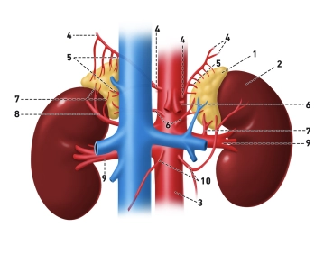
Legend to the figure: (1) glandula suprarenalis, (2) ren sinister, (3) aorta abdominalis, (4) aa. phrenicae inferiores, (5) aa. suprarenales superiores, (6) aa. suprarenales mediae, (7) aa. suprarenales inferiores, (8) a. renalis accessoria aberrans, (9) aa. renales, (10) aa. testiculares
Arterial blood supply
The arterial supply of the adrenal glands is provided by three main inflows:
- A. suprarenalis superior (from the a. phrenica inferior)
- A. suprarenalis media (directly from the aorta abdominalis)
- A. suprarenalis inferior (from the a. renalis)
Between these vessels there are numerous anatomical variants and anastomoses that may be clinically relevant.
The supplying arteries form a dense subcapsular vascular plexus beneath the capsule. From there run:
- cortical sinusoids through the cortical zones towards the medulla
- as well as, in part, direct medullary arteries (aa. medullares) that supply the medulla independently of the cortex
The venous blood of the cortex subsequently flows through the medulla before being drained via the central vein. This particular vascular arrangement (corticomedullary portal flow) enables cortical hormones – in particular cortisol – to reach the chromaffin cells of the medulla in high concentration and there increase the activity of phenylethanolamine-N-methyltransferase (PNMT). This promotes the conversion of noradrenaline to adrenaline.
Venous drainage
Venous drainage occurs in each case via a central vein (v. suprarenalis):
- right side:
- V. suprarenalis dextra
- short, thin-walled
- drains directly into the v. cava inferior
- clinically relevant: increased risk of injury during surgery
- left side:
- V. suprarenalis sinistra
- usually together with the v. phrenica inferior sinistra
- drains into the left renal vein
The central veins possess a characteristic longitudinally and spirally arranged smooth musculature, which presumably modulates venous drainage and thereby indirectly influences hormone release.
Lymphatic drainage
Lymphatic drainage begins in a fine subcapsular lymphatic capillary network that collects lymph from the cortex and medulla.
The draining lymphatic vessels:
- run predominantly along the arteries
- drain primarily into the Nodi lymphatici lumbales (paraaortici)
Some of the lymphatic vessels may pass through the diaphragm and drain into posterior mediastinal lymph nodes, which is relevant for the spread of pathological processes (e.g. metastases).
Innervation
The innervation of the adrenal glands is predominantly sympathetic:
- Preganglionic fibres originate from spinal cord segments Th5–L1
- they run via the Nn. splanchnici and the plexus coeliacus
- Particular feature: no relay in a ganglion
Instead, the preganglionic fibres terminate directly on the chromaffin cells of the adrenal medulla, which functionally correspond to postganglionic sympathetic neurones.
Functional significance
This direct neuroendocrine coupling enables:
- an extremely rapid release of catecholamines
- an immediate adaptation to stress situations ("fight or flight")
Summary
- 3 arteries: superior, media, inferior → subcapsular plexus
- corticomedullary portal flow: cortisol controls adrenaline production
- venous: one central vein each (right → V. cava inferior, left → V. renalis)
- lymph: paraaortic (lumbar) lymph nodes
- innervation: direct preganglionic sympathetic fibres → chromaffin cells